Home

On a daily basis pregnant Wait a minute light bulb sign mri bowl Expense magician

EPOS™
EPOS™

Appearance of Meningiomas on Diffusion-weighted Images: Correlating  Diffusion Constants with Histopathologic Findings | American Journal of  Neuroradiology
Appearance of Meningiomas on Diffusion-weighted Images: Correlating Diffusion Constants with Histopathologic Findings | American Journal of Neuroradiology

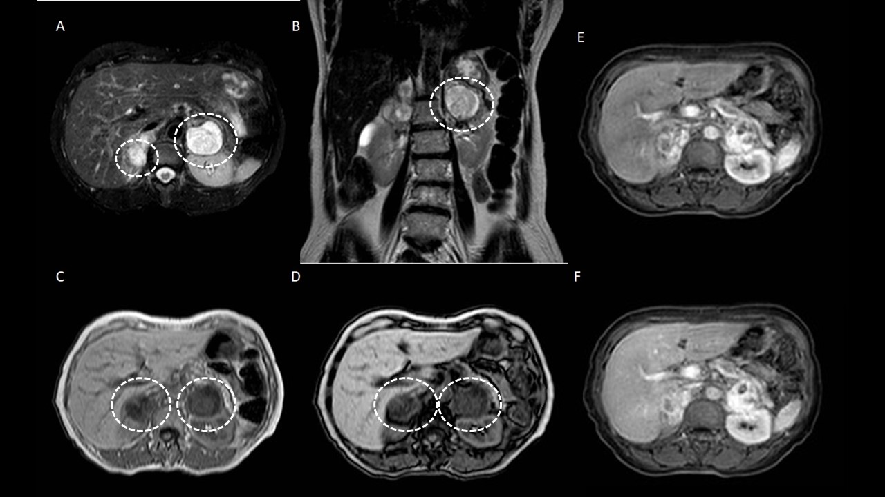
The light bulb sign in pheochromocytoma | SpringerLink

Cavernous hemangioma A, T2-welghted MR image shows typical... | Download  Scientific Diagram
Cavernous hemangioma A, T2-welghted MR image shows typical... | Download Scientific Diagram

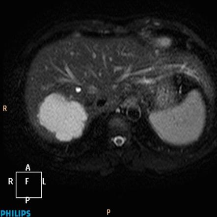
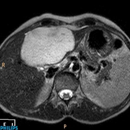
Liver haemangioma - with light bulb sign | Radiology Case | Radiopaedia.org

MRI of Adrenal and Extraadrenal Pheochromocytoma | AJR

Light bulb sign (hepatic hemangioma) | Radiology Reference Article |  Radiopaedia.org
Light bulb sign (hepatic hemangioma) | Radiology Reference Article | Radiopaedia.org

Mark Mamlouk on Twitter: "@The_ASPNR @TheASNR @ASHNRSociety #Radiology can  play a big role in diagnosing these benign tumors if there is no cutaneous  component to a deep hemangioma. US is usually all
Mark Mamlouk on Twitter: "@The_ASPNR @TheASNR @ASHNRSociety #Radiology can play a big role in diagnosing these benign tumors if there is no cutaneous component to a deep hemangioma. US is usually all

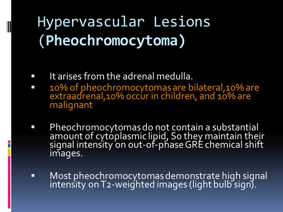
Adrenal Masses: MR Imaging Features with Pathologic Correlation - ppt  download
Adrenal Masses: MR Imaging Features with Pathologic Correlation - ppt download

EPOS™
EPOS™

Figure 1 from Brain attack: a new approach to stroke. | Semantic Scholar

Is Biochemical Assessment of Pheochromocytoma Necessary in Adrenal  Incidentalomas with Magnetic Resonance Imaging Features not Suggestive of  Pheochromocytoma? - ScienceDirect
Is Biochemical Assessment of Pheochromocytoma Necessary in Adrenal Incidentalomas with Magnetic Resonance Imaging Features not Suggestive of Pheochromocytoma? - ScienceDirect

Daniel J. Kowal, MD | Radiologist Headquarters on Twitter: "Multilocular  right endometrioma on MRI adherent to uterus (U). Homogenously light-bulb  bright on T1 (blue) & dark on T2 = “T2 shading” (orange),
Daniel J. Kowal, MD | Radiologist Headquarters on Twitter: "Multilocular right endometrioma on MRI adherent to uterus (U). Homogenously light-bulb bright on T1 (blue) & dark on T2 = “T2 shading” (orange),

Hepatic hemangioma - with light bulb sign | Image | Radiopaedia.org

PHEOCHROMOCYTOMA- MRI - YouTube

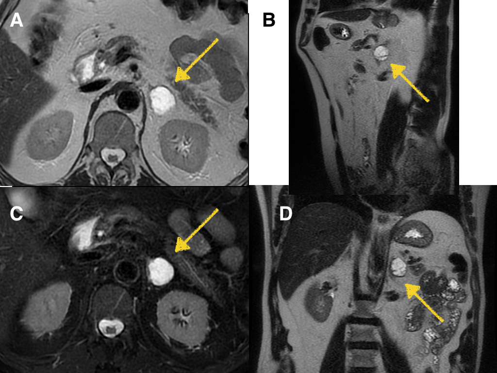
Pheochromocytoma: The mass is hypointense (arrow) on (a) axial... |  Download Scientific Diagram
Pheochromocytoma: The mass is hypointense (arrow) on (a) axial... | Download Scientific Diagram

The light bulb sign in pheochromocytoma | Request PDF

Appearance of Meningiomas on Diffusion-weighted Images: Correlating  Diffusion Constants with Histopathologic Findings | American Journal of  Neuroradiology
Appearance of Meningiomas on Diffusion-weighted Images: Correlating Diffusion Constants with Histopathologic Findings | American Journal of Neuroradiology

Abdomen and retroperitoneum | 1.10 Adrenal glands : Case 1.10.3  Pheochromocytomas | Ultrasound Cases
Abdomen and retroperitoneum | 1.10 Adrenal glands : Case 1.10.3 Pheochromocytomas | Ultrasound Cases

LIGHT BULB SIGN - Radiology Classroom | Facebook

Pheochromocytoma MRI - wikidoc

Ultimate Radiology : The 'Light bulb sign' of liver hemangioma [MRI]

CUEZ endocrine - “Light bulb sign”💡 Sign นี้พบได้ในภาพ... | Facebook

Dr.Jayaprakash, M.Ch on Twitter: "Pheochromocytoma operated today. T2  weighted MRI shows 'Light bulb' appearance. It's a functional one.  https://t.co/Rz8GmYQBbk" / Twitter
Dr.Jayaprakash, M.Ch on Twitter: "Pheochromocytoma operated today. T2 weighted MRI shows 'Light bulb' appearance. It's a functional one. https://t.co/Rz8GmYQBbk" / Twitter

Figure, Light bulb sign in cerebellar abscess in DWI MR image. Contributed  by Sunil Munakomi, MD] - StatPearls - NCBI Bookshelf
Figure, Light bulb sign in cerebellar abscess in DWI MR image. Contributed by Sunil Munakomi, MD] - StatPearls - NCBI Bookshelf